
Figure 1.
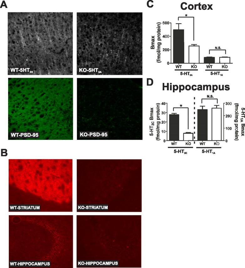
Genetic deletion of PSD-95 results in a selective loss of 5-HT2A and 5-HT2C receptors. A, 5-HT2A and PSD-95 double-label immunochemistry in medial prefrontal cortex of PSD-95wildtype and PSD-95null mice shows a large reduction in 5-HT2A receptor expression in null mice (N = 3 littermate pairs). B, 5-HT2C immunochemistry in PSD-95wildtype and PSD-95null striatum and hippocampus reveals that 5-HT2C receptor expression is almost completely abolished in the absence of PSD-95 in both striatum and hippocampus (N = 3 littermate pairs). C, Comparison of B max estimates for the 5-HT2A receptor (N = 4 littermate pairs) and the 5-HT1A receptor (N = 5 littermate pairs) in PSD-95wildtype and PSD-95null cortices. B max estimates were obtained by performing [3H]-ketanserin (5-HT2A) and [3H]-WAY100635 (5-HT1A) saturation binding on microdissected and homogenized cortical tissue. Quantitation showed an ∼40% reduction in 5-HT2A expression and no change in 5-HT1A expression in the cortices of PSD-95null mice. D, Comparison of B max estimates for the 5-HT2C receptor (N = 3; tissue from 3 animals was pooled for each measurement, for a total of 9 animals, all littermate pairs) and 5-HT1A receptor (N = 6 littermate pairs) in PSD-95wildtype and PSD-95null hippocampi. B max estimates were obtained by performing [3H]mesulergine saturation binding in the presence of 100 nm spiperone to block the vast majority of 5-HT2A receptors (5-HT2C) and [3H]-WAY100635 saturation binding (5-HT1A). Quantitation showed an almost 70% reduction in 5-HT2C expression and no change in 5-HT1A expression in hippocampus in the absence of PSD-95. All saturation binding was analyzed using nonlinear least squares fitting. B max data are presented as means ± SEM; *p < 0.05, **p < 0.01, ***p < 0.001; one-tailed unpaired t test.