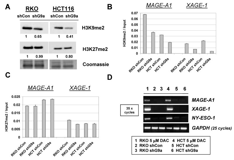
FIGURE 2.
Characterization of stable G9a knockdown human cancer cells. A. Western blot analysis of H3K9me2 and H3K27me2 levels in control shRNA and G9a shRNA expressing stable cell lines. Coomassie staining confirmed equivalent protein input, and band densitometry was performed as described in the Materials and Methods. B-C. qChIP-PCR analysis of H3K9me2 (B) and H3K27me2 (C) levels at the MAGE-A1 and XAGE-1 5′ CpG island regions. Error bars indicate + 1SD. D. RT-PCR analysis of MAGE-A1, XAGE-1, and NY-ESO-1 expression. PCR conditions and controls are the same as described in Figure 1B, and the sample key is shown below panel D.

