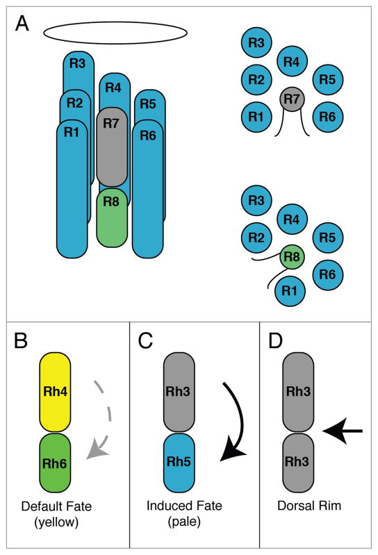
Figure 1.
Patterning of the R7 and R8 photoreceptor cells. (A) Longitudinal diagram of a Drosophila ommatidium on the left, composed of eight neuronal photoreceptor cells having rhabdomeres containing the visual pigments. The rhabdomeres of the outer photoreceptor cells (R1–R6) surround the apical and basal rhabdomeres of the inner R7 and R8 cells, respectively, as show in the cross sectional diagrams on the right. The oval is the lens. (B and C) Longitudinal diagrams of the two major types of ommatidia, yellow (B) and pale (C), expressing Rh4 and Rh6 or Rh3 and Rh5 in adjacent R7 and R8 cells. Expression of Rh6 in R8yellow cells is not dependent on the presence of an adjacent R7 cell and likely reflects a default fate (faint broken arrow). Expression of Rh5 in R8pale cells is dependent upon the presence of an adjacent Rh3-expressing R7 cell and likely reflects an inductive signal (bold arrow). (D) Ommatidia along the dorsal margin of the eye express Rh3 in both R7 and R8. The specification of ommatidia in the dorsal margin occurs from signaling events in the eye periphery and the dorsal eye that are regulated by wingless, the iroquois genes, and homothorax.16–18

