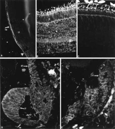
Figure 4.

Immunohistochemical detection of the FGFR-2 protein in the intact eye and the regenerating lens. Positive reaction is obvious as a fluorescent ring around the cells. (A) Presence in the lens epithelium (le) of an intact lens. lf, lens fibers. The arrowhead points to the equator. (B) Presence in all layers of intact retina. p, photoreceptors; a, amacrine cells; g, ganglion cells; op, outer plexiform layer; ip, inner plexiform layer. (C) Negative control with a section through adult retina, to show the background. (D) FGFR-2 protein is detected in the regenerating lens (le, lens epithelium; lf, differentiating lens fibers) and the dorsal iris (di, arrow) 15 days after lentectomy. (E) FGFR-2 presence in the ventral iris (vi, arrow) 15 days after lentectomy. Reaction is particularly visible in iris cells that are depigmented. At the tip of the iris (arrowhead) expression is obscured by the pigments.
