
Figure 6.
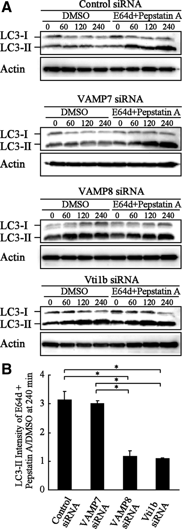
LC3-II proteins accumulated without degradation in VAMP8- and Vti1b-depleted HeLa cells. (A) HeLa cells were treated with siRNA in the same manner as described in Figure 4A. At 48 h after transfection, the cells were cultured in starved solution (EBS) with or without proteinase inhibitors for the indicated times. The cellular lysates were examined to measure the amounts of LC3-II proteins with Western blotting using anti-LC3 and actin antibodies. DMSO was used as the control. (B) Quantitative analysis of the relative intensities of the LC3-II bands (inhibitor treated/control) after 240 min in A was performed using ImageJ software. The mean values ± SD are shown from three independent experiments. *p < 0.01 by one-way ANOVA and Scheffé's posttest.