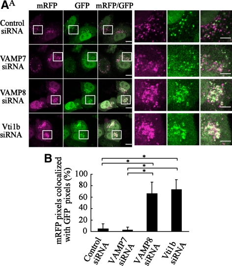
Figure 7.
Impaired degradation of LC3 proteins in autophagosomes from VAMP8- and Vti1b-depleted cells. (A) HeLa cells were transfected with siRNA for the control, VAMP7, VAMP8, and Vti1b. At 24 h after transfection, the cells were further transfected with plasmids expressing tf-LC3. After 24 h of incubation, the cells were subjected to a starved condition for 180 min and then fixed and observed with a confocal microscope. The boxed regions in the left panels are enlarged in the right panels. Bars, 10 μm (left) and 5 μm (right). (B) The colocalization frequencies of mRFP with GFP signals shown as tf-LC3 pixels in A were determined using LSM Image Browser software (Carl Zeiss) and are presented as the percentage of total number of mRFP pixels. Values are shown as the mean ± SD of >60 cell images. *p < 0.01 by one-way ANOVA and Scheffé's posttest.

