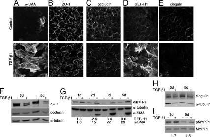
Figure 1.
TGF-β1 induces junctional disruption and GEF-H1 up-regulation in RPE cells. RPE cells were stimulated with TGF-β1 (A–E for 3 d; F–I as indicated) and processed for immunofluorescence (A–E) or immunoblot (F–H) analysis. (A–C) Samples were stained for either α-SMA (A) and ZO-1 (B), occludin (C), or GEF-H1 (D) and cingulin (E). (F–H) Immunoblots of total RPE cell extracts stimulated with TGF-β1 for the indicated time were probed with antibodies against ZO-1 and occludin (by densitometry, both proteins were decreased by >50% after 3 and 5 d of TGF-β treatment; F), GEF-H1 and α-SMA (the numbers indicate the ratios of TGF-β–treated divided by control samples obtained by densitometry; all values were normalized by those obtained for tubulin in each sample; G), cingulin (H); α-tubulin was used as loading control. (I) Immunoblot of RPE cell extracts was probed for phosphorylated (p-MYPT1) and total myosin light chain phosphatase (MYPT1) (the numbers indicate the relative increase in p-MYPT1 in TGF-β–treated samples). Shown are representative results from at least two experiments.

