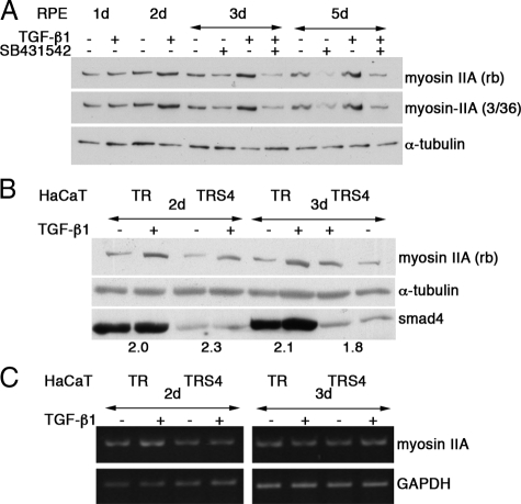
Figure 4.
TGF-β1–induced myosin-IIA up-regulation is Smad4 independent. (A) RPE cultures in the absence or presence of the ALK45 kinase inhibitor SB431542 were stimulated TGF-β1 as indicated. Immunoblots of total cell extracts are shown that were probed sequentially for myosin-IIA by using two different antibodies, a rabbit antibody (Sigma-Aldrich) or monoclonal (mouse, 3/36); α-tubulin was used as loading control. By densitometry, myosin-II was up-regulated by at least 55%. (B) HaCaT-TR-S4, a stable clone for inducible depletion of Smad4, and the parental cell line HaCaT-TR were treated with tetracycline for 2 d to reduce Smad4 expression and then stimulated with TGF-β1 for the indicated times. Total cell extracts were probed for myosin-IIA (rabbit; Sigma-Aldrich) and Smad4. The numbers indicate the ratio between TGF-β–treated and control samples for myosin-II. (C) RT-PCR analysis for myosin-IIA in control and TGF-β1–treated HaCaT-TR and HaCaT-TR-S4 cells; GAPDH served as a control to monitor RNA input. By densitometry, no significant differences were observed between control and TGF-β–treated samples.

