Figure 4.
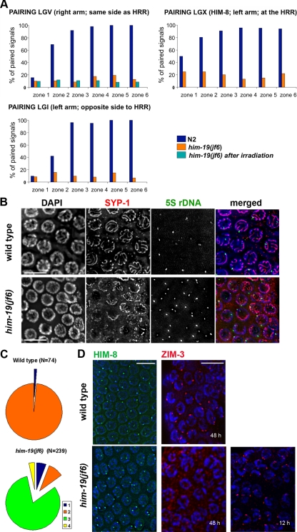
Homologous chromosome pairing is defective in the jf6 mutant. (A) Diagrams representing percentage of nuclei with homologous pairing of chromosome I, V, and X in N2, him-19(jf6), and irradiated him-19(jf6) (FISH performed 6 h after irradiation). Gonads are divided into six equal zones from the distal tip cell to diplotene (x-axes), and the percentage of pairing (y-axes) is determined by FISH probes or α-HIM-8 antibody. Levels of pairing in him-19(jf6) do not rise significantly above those in mitosis. (B) him-19(jf6) shows nonhomologous synapsis. Late pachytene nuclei are first stained with the α-SYP-1 antibody and subsequently tested by FISH with the 5S ribosomal locus probe. In him-19(jf6), a large portion of unpaired FISH signals is associated with SYP-1, indicating nonhomologous synapsis or polymerization on unpaired chromosomes. Bars, 10 μm. (C) Quantitative scoring of the 5S FISH signal in association with SYP-1 stretches in pachytene nuclei. The categories were as follows: 1 (blue), unpaired FISH signals with no association with SYP-1; 2 (orange), paired FISH signals in association with SYP-1; 3 (green), unpaired FISH signals in association with SYP-1; and 4 (yellow), paired FISH with no association with SYP-1. In the him-19(jf6) mutant, the most abundant category is the unpaired FISH signal in association with SYP-1. This either indicates frequent occurrence of nonhomologous synapsis or SYP-1 polymerization along unpaired chromosomes. (D) Immunostaining of HIM-8 (green) and ZIM-3 (red) in wild-type and him-19(jf6) pachytene nuclei. HIM-8 localizes in him-19 gonads in wild-type intensity. However, two signals are often detected in the mutant gonads indicating that the homologous pairing of the X chromosome is impaired. Two foci of ZIM-3, corresponding to the paired chromosome I set and chromosome IV set, are usually detected in wild-type TZ and early pachytene. In the him-19(jf6) background, ZIM-3 foci are detected 12 h post-L4 but not 48 h post-L4. Bars, 10 μm.

