Figure 2.
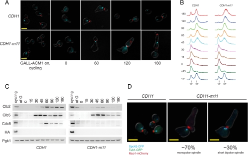
CDH1-m11 results in a first-cycle arrest with a heterogeneous spindle pole body phenotype. (A) CDH1-m11 or CDH1 cells (both GALL-HA-ACM1) were arrested in G1 with α-factor, depleted of HA-Acm1, and synchronously released. Fluorescence microscopy of Myo-mCherry (red) marking the bud neck and Tub1-CFP (cyan) were taken at the indicated time points after release from α-factor. CDH1-m11 cells multiply bud as indicated by multiple Myo1 rings. Tubulin signal varies in appearance from a point to a short bar, but elongated spindles are not observed. Bar, 5 μm. (B) Bulk DNA flow cytometry of cells as described in A. (C) Immunoblots of cells as described in A detecting the indicated proteins. Pgk1, loading control. (D) Fluorescence microscopy for Spc42-CFP (cyan) marking the SPB, Tub1-GFP (green), and Myo1-mCherry (red); 30% of CDH1-m11 cells form bipolar spindles, as indicated by two separate Spc42 dots connected by intervening tubulin-GFP. Images taken 180 min after release. Bars, 5 μm.

