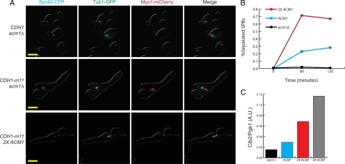
Figure 6.
ACM1 gene dosage modulates the CDH1-m11 strain spindle pole body phenotype. (A) Fluorescence microscopy for Spc42-CFP (cyan), Tub1-GFP (green), and Myo1-mCherry (red). In CDH1-m11 acm1 cells, tubulin can be seen emanating from SPBs, but separated SPBs are not observed. 2XACM1 CDH1-m11 cells can separate spindle pole bodies and form bipolar spindles. Strains were treated as described in Figure 2 and kept alive with GALL-ACM1 expression, which was shutoff in α-factor. Images were taken 180 min after release. Bar, 5 μm. (B) Percentage of synchronized acm1, wild type (1X ACM1), and 2X ACM1 cells, all with CDH1-m11, displaying separated spindle pole bodies at indicated time points. (C) Clb2 levels for indicated genotypes, all with endogenous CDH1-m11, at 60 min after release from α-factor, standardized to Pgk1 loading control.

