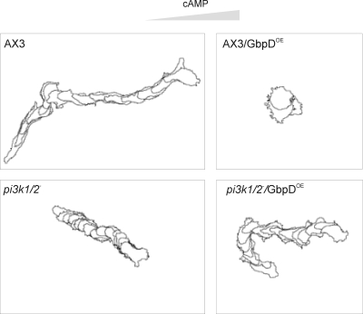
Figure 2.
Chemotaxis assay. To analyze the chemotaxis behavior of wild-type (AX3), pi3k1/2−, AX3/GbpDOE and pi3k−/GbpDOE strains, cells were starved and pulsed for 6–8 h, resuspended in PB, and monitored by phase-contrast microscopy. Cells were stimulated with a micropipette containing 10−4 M cAMP, from the right. The contours of the cells are shown at 1-min interval for AX3, pi3k1/2−, and pi3k1/2−/GbpDOE and at 5-min intervals for AX3/GbpDOE for a total of 15 min.

