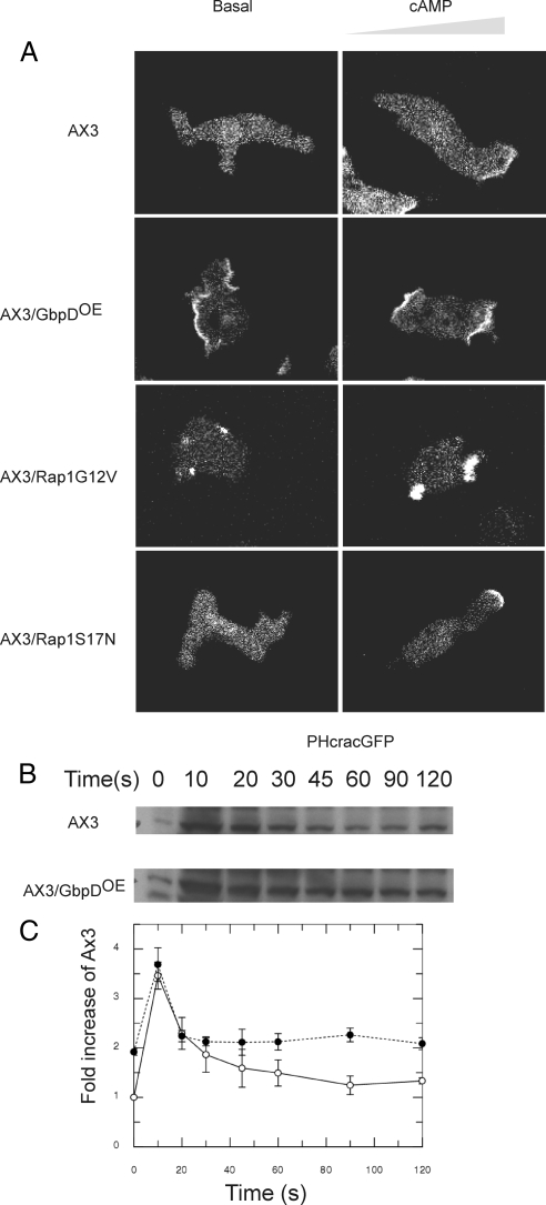
Figure 3.
Effect of GbpD expression on PI3K activity. (A) To investigate the effect of GbpDOE on PIP3 levels, the PIP3 detector PHcracGFP was expressed in wild-type, GbpDOE, RapG12VOE and RapS17NOE cells. Confocal images are shown for unstimulated cells (left) and for cells stimulated with cAMP from a micropipette on the right (right). Movies 1 and 2 for wild-type and GbpDOE cells, respectively, are available as Supplementary Information. (B) PKB phosphorylation in wild-type and GbpDOE cells. Cells were cAMP pulsed and then stimulated with 1 μM cAMP. Samples were removed at the times indicated and lysed directly into SDS loading buffer. Samples were subjected to SDS-PAGE and analyzed by Western blotting by probing with a phospho-threonine–specific antibody. The indicated ∼51-kDa protein corresponds to the phosphorylation of PKB/Akt. The blot shown is representative of three independent experiments. (C) Quantification of PKB phosphorylation for AX3 (○) and GbpDOE (•) cells. Data are presented as fold increase relative to AX3 cells before stimulation.

