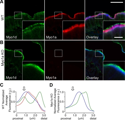
Figure 4.
Myo1a and Myo1d exhibit differential localization within the BB. (A) Confocal images of adult WT mouse small intestine frozen sections stained for Myo1d (green, C13 antibody), Myo1a (red), and F-actin (blue). In the BB, Myo1d occupies microvillar tips and the terminal web, whereas Myo1a localizes along the length of microvilli. (B) KO mouse sections stained in an identical manner reveal that Myo1d is found along the length of microvilli in the absence of Myo1a. Myo1d still occupies microvillar tips, but redistributes from lateral plasma membrane and the terminal web. (C and D) Plots show the average pixel intensity along the microvillar axis from proximal (base) to distal (tip) for Myo1d (green), Myo1a (red), and phalloidin (blue) fluorescence signals. The arrow indicates the position of the terminal web. Representative micrographs of “straightened” BBs used to create these plots are shown in Supplemental Figure 3. Bar, 20 μm; inset bar, 5 μm; bars serve as calibration for A and B.

