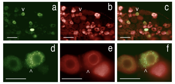
Figure 14.
Demonstration of transport to C-type nociceptor cells in dorsal root ganglion. Section of rat L5 dorsal root ganglia showing retrogradely transported FITC combined with immunohistochemistry for the specific C-fiber nociceptor marker, Peripherin. (a) - Retrogradely transported FITC, (b) - the same field as in (a) showing cells that are immuno-positive for peripherin, (c) - overlay of (a) and (b) showing FITC is present in the nociceptors (v - same neuron in a, b, &c). There are 26 cells seen with FITC in (a) versus 59 cells with peripherin in (b) & (c). (d) - Two sensory neurons containing retrogradely transported FITC, a third neuron is unlabeled, (e) - the same field as in (d) showing that all three neurons are positive for Peripherin, (f) - overlay of (d) and (e) (^ - same neuron in d, e, &f). Scale bars (a-c) = 150 μm, (d-f) = 40 μm.

