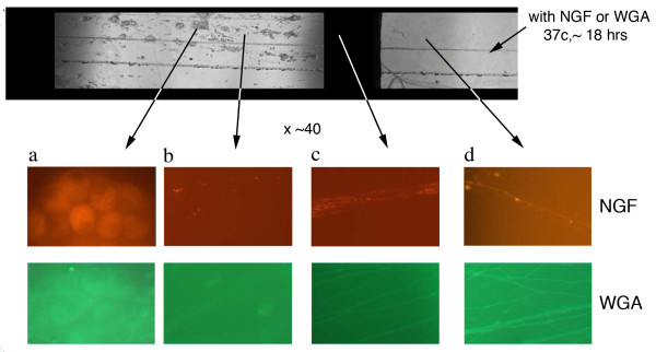
Figure 4.
Axonal Transport of NGF and WGA in Campenot Chambers. Top panel shows a transmission image of the central and side compartments of a Campenot chamber. WGA-FITC or NGF-Texas Red were added to the side compartment and left overnight. At higher magnification, the "red series" of panels are fluorescent images obtained from the arrowed areas of the chamber following NGF-TR administration. Similarly, the "green" series of panels shows fluorescent images taken of the chambers following WGA-FITC administration. The left most panels in both series show uptake of fluorescence in cell bodies derived from the axonal transport of these labeled ATFs.

