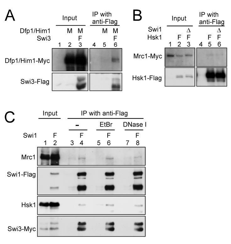
Figure 1. Interaction of Hsk1-Dfp1/Him1 with Swi3 and Mrc1.
(A) Co-immunoprecipitation of Dfp1/Him1 with Swi3. (B) Co-immunoprecipitation of Mrc1 with Hsk1. The extracts were prepared from the strains indicated, and immunoprecipitation with anti-Flag antibody was performed as described in “Experimental procedures” (lanes 4-6 in (A) and lanes 4-6 in (B)). Input (lanes 1-3 in (A) and lanes 1-3 in (B)) represents 2.5 % of the extracts used for the immunoprecipitation. Western blotting analyses were conducted using the antibodies against the tag as indicated. (A) lanes 1 and 4, YM71; lanes 2 and 5, EN3404; lanes 3 and 6, SH1007. (B) lanes 1 and 4, KT2791; lanes 2 and 5, MS404; lanes 3 and 6, MS405. (C) Co-immunoprecipitation of Hsk1 with Swi1 from EtBr or DNaseI treated extracts. The extracts from the strains indicated were treated with either EtBr (50 μg/ml) or DNase I (0.35 U/μl) for 15 min at 0°C, followed by immunoprecipitation with anti-Flag antibody (lanes 3-8). Input (lanes 1 and 2) represents 1.7 % of the extracts used for the immunoprecipitation. Western blotting analyses were conducted using the antibodies indicated. Lanes 1, 3, 5 and 7, SH0987; lanes 2, 4, 6 and 8, SH1232. Genotypes are indicated above each lane with the abbreviation as follows. M, Myc-tagged; F, Flag-tagged; Δ, deletion.

