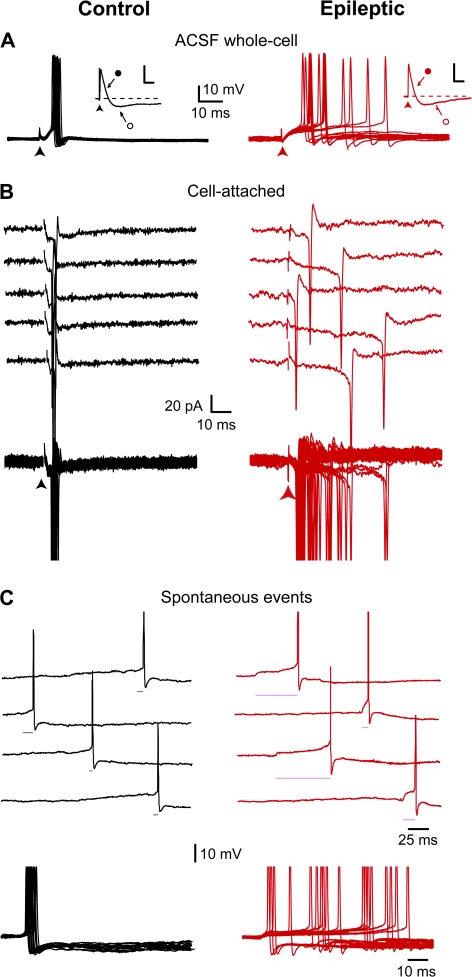
Figure 5.
Reduced temporal precision of EPSP-spike coupling in physiological conditions. These experiments were performed in ACSF, in the absence of GABA and NMDA receptor blockers. (A) Superimposed suprathreshold EPSPs recorded in a granule cell from control (black traces) and epileptic (red traces) rats. Note time-locked (SD = 1.0 ms) and jittered spikes (SD = 13.4 ms) in control and epileptic rats, respectively. When EPSP (close circle) is followed by a di-synaptic IPSP (open circle) this shortens EPSP decay and prevents the generation of action potentials both in DGCs from control (black inset) and epileptic (red inset, scale bars: 2 mV, 50 ms) rats. (B) Individuals (top) and superimposed (bottom) loose cell-attached recordings of spiking in response to electrical stimulations in a granule cell from control (black traces) and epileptic (red traces) rats. Note time-locked (SD = 0.6 ms) and jittered spikes (SD = 9.9 ms) in control and epileptic rats, respectively. (C) Continuous recordings of spontaneous suprathreshold EPSPs (top) and superimposed spontaneous suprathreshold EPSPs (bottom, aligned on the rising phase) in a granule cell from control (black traces) and epileptic (red traces) rats. Note that spontaneous EPSPs always trigger spikes from their rising phase in the granule cell from control rat, whereas in the granule cell from epileptic rat spikes are generated either from the rising phase or during the long-lasting plateau of spontaneous EPSPs; EPSP-spike latency is indicated by a horizontal line.

