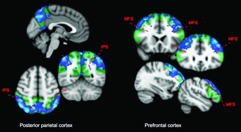
Figure 4.
Prefrontal and posterior-parietal subregions contributing to resting state connectivity with the posterior cerebellum. These connectivity maps were generated by taking the first Eigen time series from the cerebellar supramodal zone, and identifying voxels in the prefrontal and posterior-parietal cortex which correlated with it. Hence, they indicate which voxels within the cerebral–cortical regions contribute to the resting state correlation with the posterior cerebellum. Green tinted regions show the extent of the cortical regions. Blue statistic maps indicate group Z-scores thresholded at Z > 1.6 (equivalent of P < 0.05 uncorrected). Note that even at this low threshold, the parietal correlation map is limited to the inferior parietal lobule and medial parietal cortex—there is a clear boundary between significant and nonsignificant correlation at the intraparietal sulcus (especially clear on axial view). The prefrontal correlation map does not extend into the inferior frontal gyrus; a boundary at the inferior frontal sulcus is clearly visible in the coronal views. MFS, medial frontal sulcus; IPS, intraparietal sulcus.

