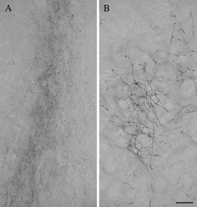
Figure 10.
The micrographs illustrate the morphology of axons and terminals labeled by an injection of BDA in the temporal cortex. In the pulvinar nucleus (A), the labeled axons are of fine caliber and give rise to small boutons. In the PT (B), axons labeled from the same cortical injection site are thicker and give rise to larger boutons. Scale bar = 30 μm and applies to both panels.

