
Figure 2.
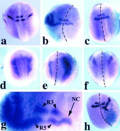
Expression of Krox-20 (a–c, g, and h) and slug (d–f) in Xenopus embryos (stage 20–25), as seen by whole-mount in situ hybridization. The embryos are viewed from the dorsal side, and anterior is to the top. Krox-20 expression in a control embryo (a) is in two stripes (rhombomeres 3 and 5) in the hindbrain and a stream of neural crest cells adjacent to rhombomere 5. Xwnt-3a-injected embryo (b); in the injected half (note the turquoise β-gal staining in the left half) the stream of Krox-20-expressing cells is dramatically expanded while still arising from a zone adjacent to rhombomere 5. In an Xwnt-8-injected embryo (c), the pattern of expression of Krox-20 is unaffected. Slug expression in a control embryo (d). Xwnt-3a-injected embryo (e) shows an enlargement of the domain of expression of slug within the injected right half (see β-gal staining). An Xwnt-8-injected embryo (f) presents a pattern of slug expression in the injected half (right side, β-gal staining) comparable to the uninjected half. (g) Higher magnification of the hindbrain region of the embryo presented in b. Rhombomere 3 (R3) and rhombomere 5 (R5) expression of Krox-20 is not affected; NC indicates stream of neural crest. CS2+Xwnt-3a-injected embryo (h) presents an expanded domain of expression of Krox-20 in the injected half (see β-gal staining). The dotted lines indicate the dorsal midline. Among the embryos injected in the animal pole region, 30% showed some level of axis duplication when injected with Wnt-1, Xwnt-3a, or Xwnt-8 RNAs.