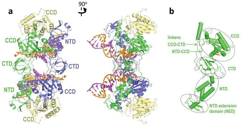
Figure 1. Architecture of the PFV intasome.
a, Views along (left) and perpendicular (right) to the crystallographic two-fold axis. The inner subunits of the IN tetramer, engaged with viral DNA, are blue and green; outer IN chains are yellow. The reactive and non-transferred DNA strands are magenta and orange, respectively. Side chains of Asp128, Asp185 and Glu221 active site residues are red sticks; gray spheres are Zn atoms. Locations of the canonical IN domains (NTD, CCD and CTD) are indicated; b, inner (green) IN chain with domains and linkers indicated. The orientation is the same as in the right panel of a.

