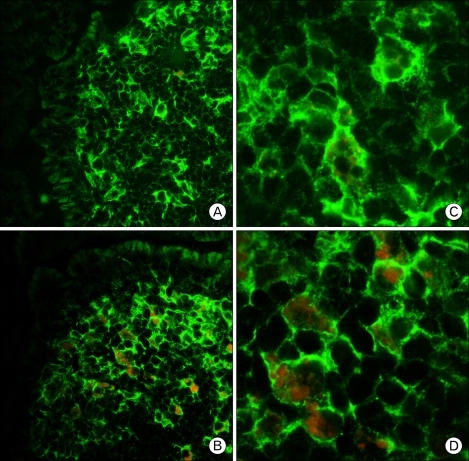
Figure 4.
Localization of CCL3 in the Peyer's patches. Frozen sections of Peyer's patches were doubly stained with antibodies against CD11c (green) in combination with anti-CCL3 (red). Panels (A) and (C) display the controls, and panels (B) and (D) display the ginsan treatment. (A, B ×600; C, D ×1,800).

