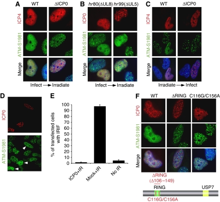
Figure 1.
Expression of ICP0 prevents accumulation of DNA repair proteins at IRIF. (A) HeLa cells were infected with WT or ICP0-null HSV-1 at an MOI of 5 for 2 h and treated with 10 Gy IR. Cells were fixed 1 h post IR, stained for ICP4 and assessed for localization of proteins reacting with an antibody generated to phosphorylated ATM. Nuclei were stained with DAPI as shown in the merged image. (B) HeLa cells were infected with ΔUL8 or ΔUL5 mutants of HSV-1 at an MOI of 5 and then treated with 10 Gy IR. Cells were fixed 1 h post IR, and stained with antibodies to ICP0 and phosphorylated ATM. (C) ICP0 can disrupt pre-established IRIF. Uninfected HeLa cells were irradiated at 10 Gy and infected 1 h post IR with WT or ICP0-null HSV-1 at an MOI of 5 for 2 h. Cells were fixed 2 h post infection and stained with antibodies to ICP4 and phosphorylated ATM. (D) HeLa cells were transfected with an ICP0 expression plasmid for 16 h and then treated with 10 Gy IR. Cells were fixed 1 h post IR, and stained with antibodies to ICP0 and phosphorylated ATM. (E) Transfected HeLa cells (200) were scored for the localization at IRIF of proteins reacting with phosphorylated ATM antibody. Data are represented as mean±s.e.m. (F) HeLa cells were transfected with WT or RING mutant ICP0 expression plasmids for 16 h and then treated with 10 Gy IR. Cells were fixed 1 h post IR, and stained with antibodies to ICP0 and phosphorylated ATM.

