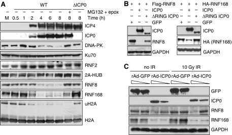
Figure 4.
ICP0 induces degradation of RNF8 and RNF168. (A) HeLa cells were infected with WT or ΔICP0 HSV-1 at an MOI of 5 and lysates prepared for immunoblotting over a timecourse of infection. Degradation of RNF168 and loss of ubiquitinated H2A were detectable at 2 h post infection, whereas DNA-PKcs and RNF8 degradation were detectable at 4 h post infection. Levels of all degradation substrates were rescued by proteasome inhibitors added at 2 h (compare levels of RNF8 and RNF168 at 2 h to levels at 8 h in the presence of MG132 and epox). Ku70 served as a loading control. (B) HeLa cells were transfected with Flag-RNF8 or HA-RNF168 and either ICP0 WT or the ΔRING mutant in a ratio of 3:1, where ICP0 was in excess. Cells were collected at 20 h post transfection, lysates were prepared for immunoblotting and assessed for levels of RNF8 and RNF168. GAPDH was used as a loading control. (C) HeLa cells were infected with recombinant adenoviruses expressing either GFP or ICP0, plus recombinant adenovirus expressing the tet activator (1:1) at a total MOI of 50 or 10. Cells were irradiated 23 h.p.i. and then collected 1 h post IR. Lysates were analysed by immunoblotting and assessed for levels of RNF8 and RNF168. GAPDH was used as a loading control.

