
Fig. 7.
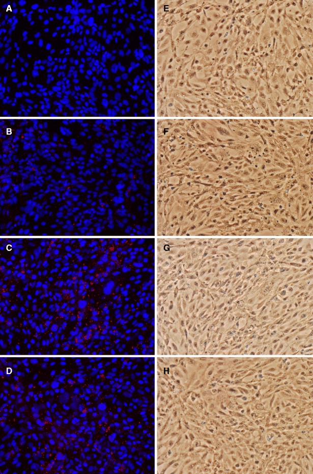
Microscopic observation of hCMEC/D3 cells incubated with phage. A–D. Fluorescence microscopy image of phage labeled with alexa-568 (red) and DAPI-stained cell nuclei (blue). A. No phage. B. Control phage RVR. C. Selected phage GLA. D. Selected phage GYR. E–H. Light microscopy image of the same view on the cell monolayer as the fluorescence image to the left.