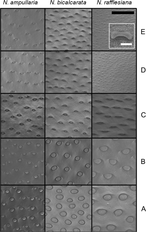
Fig. 2.
Scanning electron microscope (SEM) images comparing the inner pitcher surface at zones A through E for N. ampullaria, N. bicalcarata, and N. rafflesiana. Inset outlined in white for N. rafflesiana shows individual lunate cell. Note pale epicuticular wax crystals on surface. Black bar=0.5 mm; white bar=25 μm.

