Figure 6.


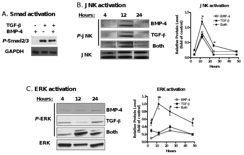
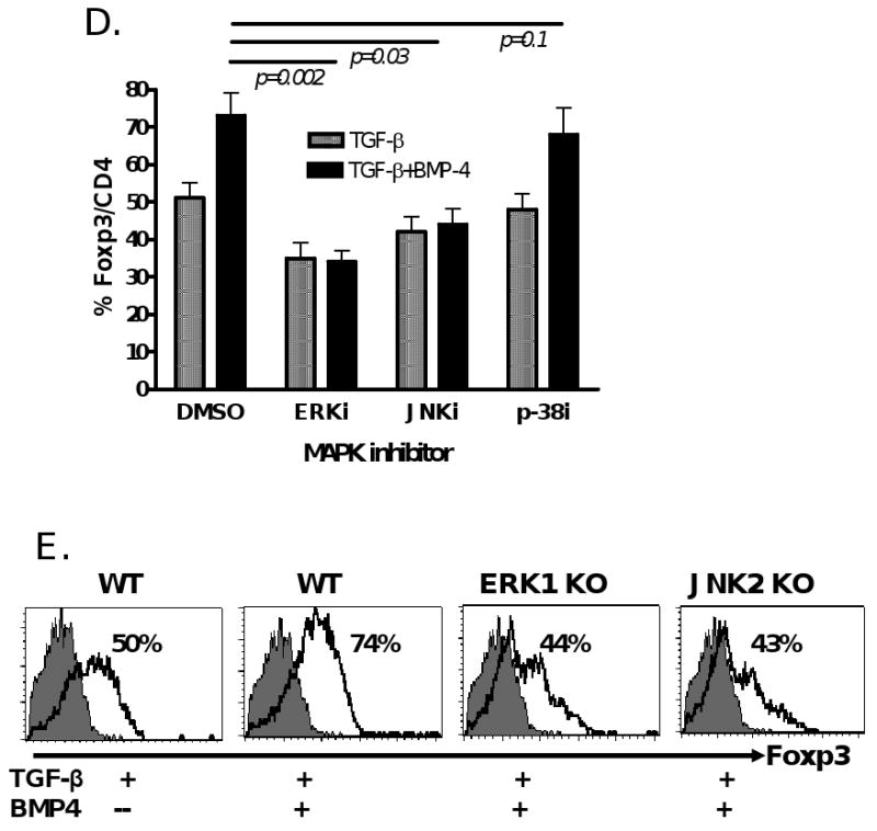
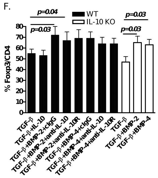
BMP2/4 on enhancement of TGF-β-induced CD4+Foxp3+ Treg development involves MAPK activation. (A) Naïve CD4+ cells were TCR (anti-CD3/CD28 coated bead 1:5) stimulated with or without BMP-4 ± TGF-β for 16 hours. Activated Smad2/3 expression was determined by western blot. These cells were stimulated with TCR in the presence of BMP-4, TGF-β or both (BMP-4+TGF-β) in various time-points as indicated and the levels of activated JNK (B) and ERK (C) were determined by western blot. (D) Foxp3 expression by TCR-stimulated CD4+ cells cultured with BMP-4 + TGF-β in the presence or absence of MAPK inhibitors. (E) Foxp3 expression by TCR-stimulated CD4+ cells cultured with BMP-4 ± TGF-β in ERK1 KO, JNK2 KO and littermate control mice for 4 days. (F) Foxp3 expression on CD4+ cells stimulated with TCR with BMP-2/4 ± TGF-β in IL-10 KO and littermate control mice for 4 days. In some experiments with wild type control mice, anti-IL-10, anti-IL-10R or control IgG were added to cultures. Data show mean ± SEM of three separate experiments or represent at least three independent experiments.

