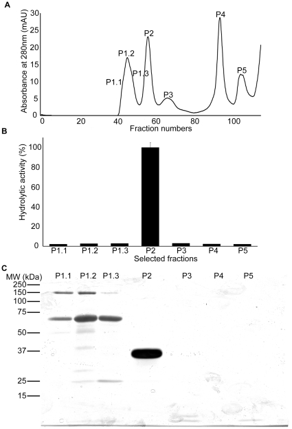
Figure 2. Purification of rhinocerase from rotofor separated venom fractions.
A. Superdex 75 gel filtration chromatogram obtained during the elution of proteins from rotofor separated venom fractions 3 and 4. The peaks were numbered at the particular fractions which were used for analysis by SDS-PAGE and for analysis of the serine protease activity. B. 100 µl of the selected fractions shown in figure A were used to measure serine protease activity using Arg-AMC fluorescent substrate. Each bar shows the mean ± S.D. (n = 3). The hydrolytic activity measured for P2 was taken as 100%. C. 100 µl of the selected fractions indicated in figure A were analysed by SDS-PAGE (10%) and silver stained.

