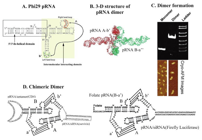
FIG. 1.
Sketch of sequence and structure of pRNA chimeras. (A) φ29 pRNA sequence and secondary structure. The right- and left-hand loops are circled in orange and green, respectively. The double-stranded helical domain on the 5′/3′ ends is framed, and the domain for dimer formation is shaded. The curved line points to the two interacting loops. (B) Three-dimensional structure of pRNA dimer. (C) Native polyacrylamide gel showing monomer and dimer of the pRNA chimeras exhibiting different migration rates. Below the gel are cryo-AFM images of ′29 pRNA monomer and dimer. The colors reflect the thickness and height of the molecule; the brighter the color, the thicker or taller the molecule. (D) Design of chimeric pRNA dimers harboring foreign moieties (see Nomenclature of RNA Subunits, under Results).

