FIG. 9.
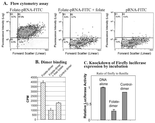
Specific delivery of chimeric pRNA/siRNA by folate-pRNA. (A) Flow cytometry analyses of the binding of FITC-labeled folate-pRNA to KB cells. Left: Cells were incubated with folate-pRNA labeled with FITC. Middle: Cells were preincubated with free folate, which served as a blocking agent to compete with folate-pRNA for binding to the receptor. Right: Binding was also tested using folate-free pRNA labeled with FITC as a negative control. The percentages of FITC-positive cells are shown in the top right quadrants. (B) Specific binding of folate-pRNA dimer to KB cells. After incubation of cells with the [3H]folate-pRNA dimer in the presence (middle column) or absence (left column) of free folate, cells were isolated and subjected to scintillation counting. The right-hand column represent 3H-labeled dimer without folate labeling as a negative control. (C) In a knockdown assay by incubation, folate-chimeric dimer complex containing pRNA(B-a′)/folate and pRNA(A-b′)/siRNA(firefly) was incubated with KB cells for 3 hr to allow the binding and entry of RNA. The luciferase level was measured the next day in the dual reporter system. The control dimer was identical to the folate dimer except for its lack of folate labeling.

