
Figure 3. Effects of PHT-427 in cells.
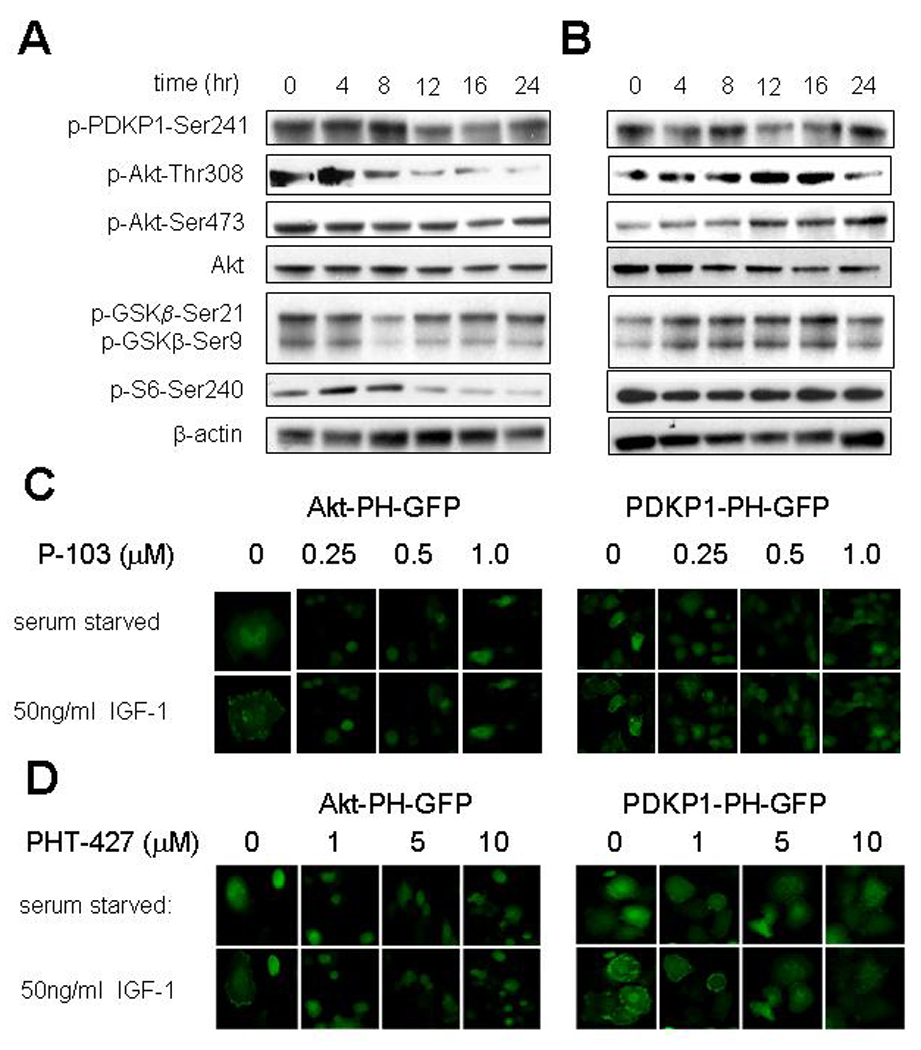
Western blots of Akt activity measured by phospho-Ser473-Akt and PDPK1 activity by phospho-Ser241-PDPK1 and activation of selected downstream targets. β-Actin was used as a loading control. A, BxPC-3 pancreatic cancer cells exposed to 10 µM PHT-427 in media with 10% fetal bovine serum (FBS) for various times; B, MiaPaCa-2 pancreatic cancer cells exposed to 10 µM PHT-427 in media with 10% FBS for various times; C, Panc-1 pancreatic cancer cells stably transfected with Akt-PH domain-GFP or PDKP-1-PH domain-GFP were cultured in serum free medium for 16 hr, exposed to the PtdIns-3-K inhibitor P-103 at different concentrations for 4 hr, and then stimulated with IGF-1 50 ng/ml for 20 min or no stimulation. Cellular fluorescence was measured with an IN Cell Analyzer 1000; D, similar studies with Akt-PH domain-GFP or PDKP-1-PH domain-GFP Panc-1 cells exposed to PHT-427 at different concentrations.