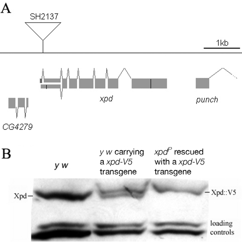
Figure 1. xpd gene and alleles.
(A) Map of the xpd genomic region. Known transcripts and the P-element insertion (triangle) in xpd are shown (figure based on the information provided by www.flybase.org). Boxes depict exons, connecting lines introns of the different transcripts. The alternative exons at the 5′ end of xpd are visualized by split boxes, with the two different translational starts shown as black vertical bars. The position of the translational stop codon in the last exon is also indicated by a black bar. (B) xpd-V5 rescues the xpdP mutant phenotype. Total fly extracts were analyzed by Western blotting with anti-Drosophila Xpd antibodies. xpd-V5 transgenic lines express similar levels of exogenous and endogenous Xpd. Unidentified cross reacting bands serve as loading controls. In xpdP flies rescued with xpd-V5, only Xpd::V5 is detected.

