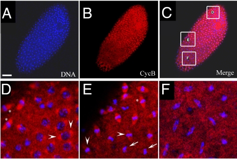
Figure 7. Cyclin B distribution in asynchronous mitoses of xpdeE embryos.
(A–F) The mutant cycle 11 embryo was stained with Hoechst (blue) and anti-CycB (red). Scale bar represents 50 µm. Nuclear divisions (A) and CycB degradation (B) are not synchronized throughout the embryo. However, the latter takes place at the appropriate cell cycle stage. (D–F) show regions from (C) at higher magnification. Arrowheads in (D) show CycB localized to spindle regions of prophase nuclei. “*” in (D,E) show CycB localized to spindle regions and DNA regions of early metaphase nuclei. Arrowheads in (E) show levels of CycB drop in spindle regions as nuclei progress through metaphase. Arrows in (E) show no localization of CycB to spindle regions in late metaphase/early anaphase nuclei. The same is the case in the anaphase nuclei shown in (F).

