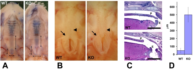
Figure 2. Wnt1-Ate1 mice have palate defects.
(A) Observation of the roof of the mouth of control (WT) and Wnt1-Ate1 (KO) newborn pups at P0 shows a shorter soft palate area (longer vertical arrow on the right in each image) and an enlarged nasopharynx entrance (two shorter perpendicular arrows on the left and bottom in each image). Heads have been contrasted with trypan blue dye for better observation. 5 wild-type and 5 Wnt1-Ate1 pups were analyzed. (B) Closer view of the nasopharynx entrance (arrow) and the surrounding area in a control (WT) and aWnt1-Ate1 (KO) newborn pup at P0 with the missing soft palate (arrowhead). (C) Sagittal sections through the palate areas of the control (WT) and Wnt1-Ate1 (KO) newborn pups at P0. While in control the soft palate (P) reaches all the way to the epiglottis (E), in the mutant the palate is shorter and leaves a large gap of exposed tissue at the back of the throat. Scale bar, 1 mm. 4 wild-type and 5 Wnt1-Ate1 pups were analyzed. (D) Quantification of the distance from the end of the palate to the epiglottis in all the examined pups at P0 (4 wild-type and 5 mutants). In wild-type, the average distance was 48.6+/−28.9 (SEM), and in the mutants this number increased over 10-fold to 501.0+/−93.7 (SEM), indicating a significant overall shortening of the tissue of the soft palate.

