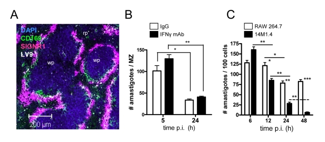
Figure 1. Splenic marginal zone macrophages and marginal metallophilic macrophages control L. donovani by an IFN-γ independent mechanism.
(A) L. donovani amastigotes are found predominantly in MMM and MZM after intravenous injection (purple, SIGNR1; green CD169; white, L. donovani; blue DAPI). (B) C57BL/6 mice were treated with 0.5mg XMG 1.6 anti-IFN-γ IgG (black bars) or control IgG (open bars) and 2h later infected with L. donovani. At 5h and 24h p.i., the number of parasites per MZ area was determined. (C) 14M1.4 cells (black bars) and RAW 264.7 cells (open bars) were infected with L. donovani (MOI 10∶1) and at indicated times p.i., parasite numbers per 100 macrophages were determined by fluorescence microscopy. *, p<0.05, **, p<0.01 between bars indicated as indicated. Scale bar; 200µm.

