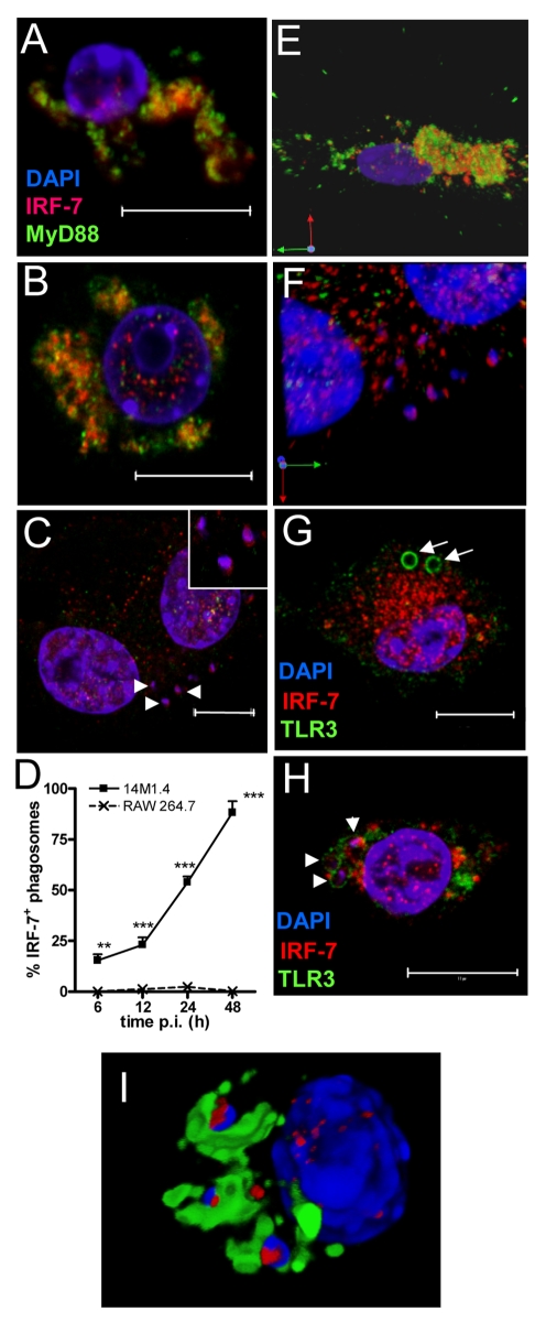
Figure 3. IRF-7 is recruited to Leishmania phagosomes.
Untreated (A), poly(I:C)-treated (B) and 48h L. donovani-infected (C) 14M1.4 cells were stained for IRF-7 (red) and MyD88 (green). Cells were counterstained with DAPI (blue) and analysed by confocal microscopy. Single confocal slices are shown. Amastigotes are indicated with arrowheads and enlarged in inset in (C). (D) Frequency of IRF-7+ phagosomes was scored at time intervals shown in infected 14M1.4 and RAW264.7 cells. (E, F) Snap shots from Video S1 and S2, showing IRF-7-MyD88 association in untreated 14M1.4 cells (E) and phagosomal ‘capping’ of IRF-7 in Leishmania-infected 14M1.4 cells (F) (red, IRF-7; green, MyD88; blue, DAPI.). (G) 14M1.4 cells stimulated with poly (I:C) and allowed to phagocytose latex beads (arrow), stained for IRF-7 (red) and TLR3 (green). (H) 14M1.4 cells infected with L. donovani and stained for TLR3 (green) and IRF-7 (red). Scale bars; 10µm. (I) Snap shot from Video S3, showing IRF-7 and LAMP1 staining in 24h Leishmania-infected 14M1.4 cells (red, IRF-7; green, MyD88; blue, DAPI).

