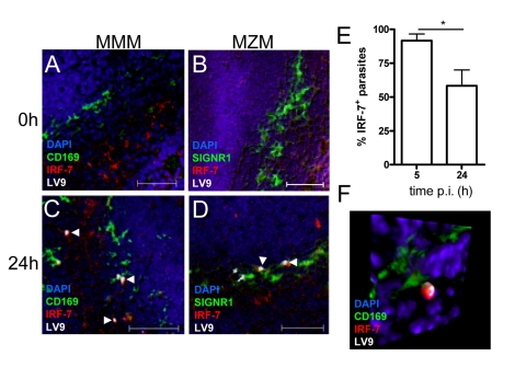
Figure 4. IRF-7 is induced in MZM and MM in situ.
(A–D) Induction of IRF-7 in MMM and MZM. Naïve (A,B) and C57BL/6 mice infected for 24h (C, D) were stained for CD169 (A, C; green) or SIGNR1 (B, D; green), IRF-7 (red) and L. donovani (white). (E) The percentage of IRF-7+ parasite-containing phagosomes was quantified by counting 100 amastigotes/section (n = 3) and calculating the proportion of parasites with IRF-7 accumulation. *, p<0.05 (F) Snap shot from Video S4, showing infected MMM with phagosomal localisation of IRF-7 (CD169+ green, IRF-7, red; LV9, white; DAPI, blue). Scale bars; 50µm.

