Figure 1.
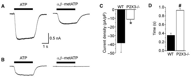
Stimulation of dissociated gastric sensory neurons with ATP and α,β-metATP. (A) Current responses during superfusion with 30 μmol L−1 of ATP (left) and 30 μmol L−1 of α,β-metATP (right) from a wildtype animal. (B) The corresponding responses from a gastric neuron of a P2X3−/− mouse. The peak current density (C) and time to peak (D) in response to 30 μmol L−1 of ATP are summarized for wildtype (black bars; n = 15) and P2X3−/− mice (white bars; n = 22). *P < 0.05; P < 0.01.

