Figure 4.
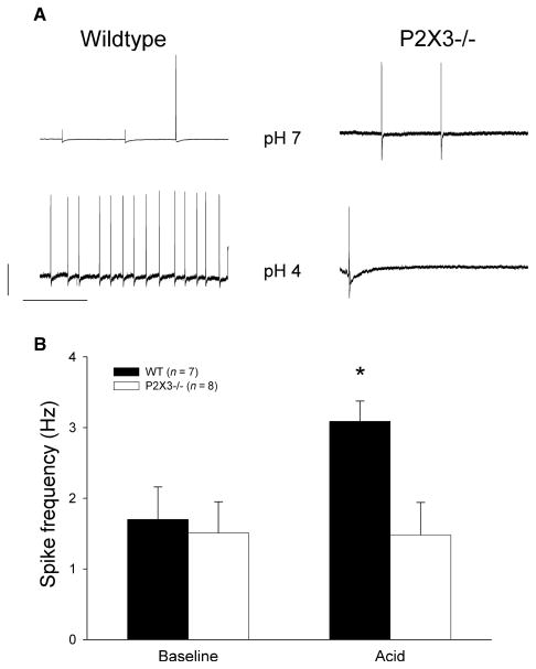
Effect of luminal acidification. (A) Representative voltage tracings with action potential firing after luminal perfusion at pH 7 (upper tracings) and pH 4 (lower tracings) for wildtype (left) and P2X3−/− mice (right). The calibration bars represent 20 mV and 1 s respectively. The data are summarized in the bar graph. (B) The average spike frequency at baseline (left side) and 3 min after decreasing the pH to 4 in wildtype animals (black bars) and P2X3−/− mice (white bars). While acidification triggered a significant increase in controls (P < 0.05), spike frequency did not change after exposure to pH 4 knockout mice.

