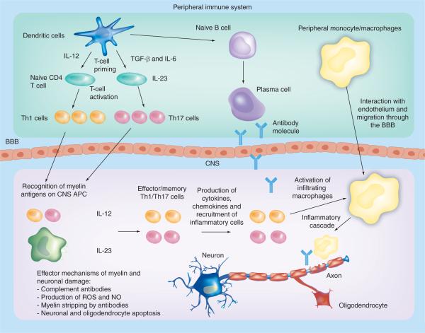
Figure 1. Pathogenesis of experimental autoimmune encephalomyelitis and multiple sclerosis.
In the peripheral immune system, APCs (mainly DCs but also B cells and macrophages) produce IL-12, TGF-β and IL-6, and prime naive CD4+ T cells during activation by interaction of the T-cell receptor with cognate antigens presented by MHC class II complexes. Activated Th1 and Th17 cells cross the BBB into the CNS. Resident APCs interact with activated myelin-specific Th1 and Th17 cells, which become reactivated (Th2 cells can also be primed by APCs). Inflammatory cytokines and chemokines are produced and an inflammatory cascade is initiated. Infiltrating macrophages become activated and these, alongside antibodies, attack the myelin sheath on neurons.
APC: Antigen-presenting cell; BBB: Blood-brain barrier; NO: Nitric oxide; ROS: Reactive oxygen species; Th: T helper.

