Fig. 4.
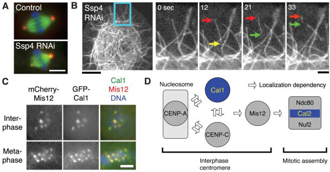
Regulation of spindle length and chromosome alignment. (A) Spindle length was altered after RNAi depletion of the novel protein Ssp4. Scale bar, 5 µm. (B) MT severing (yellow arrow) frequently occurred after Ssp4 RNAi. Severed MTs often showed treadmilling behavior (red and green arrows) and then disappeared. Scale bars, 10 µm (left), 2 µm (right). See also movie S4. (C) Previously unknown Cal1 protein localizes to the centromere (marked by mCherry-Mis12). (Localization data for other proteins are in fig. S7). Scale bar, 2 µm. (D) Model for kinetochore assembly in S2 cells based on protein localization and RNAi. (Data are in fig. S7, D to F).

