
Figure 2.
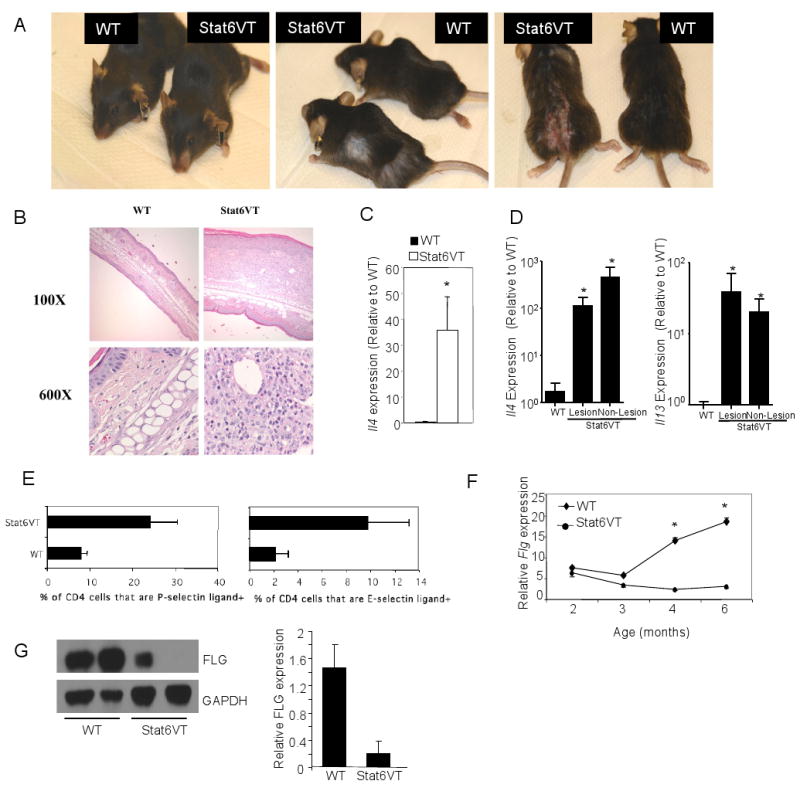
Mice expressing constitutively active Stat6 in T cells develop allergic skin inflammation. A, Photographs of Stat6VT transgenic mice and littermate controls showing areas affected by skin inflammation. B, Histological analysis of ear tissue from wild type and Stat6VT transgenic mice. Samples were fixed and stained with hematoxylin/eosin. C, RNA was isolated from skin of wild type and Stat6VT transgenic mice that did not have lesions before analysis of cytokine mRNA using qPCR. D, RNA was isolated from skin of wild type and lesional or non-lesional skin of Stat6VT transgenic mice that had active lesions before analysis of cytokine mRNA using qPCR. Results are the average ± SEM of 4-5 mice. E, Selectin ligand expression was examined on CD4+ splenic T cells using flow cytometry. Results are the average ± SD of 2-4 mice and are representative of two experiments. F, RNA was isolated from skin of wild type and Stat6VT transgenic mice that did not have lesions before analysis of Flg mRNA using qPCR. G, Immunoblots of protein extracts from epidermis of WT and Stat6VT transgenic mice for filaggrin (FLG) with GAPDH as a loading control. Bar graphs represent the average densitometry of 3-5 samples normalized to expression of GAPDH. Results in panels C, D and F are the average ± SEM of 5-9 mice. *, significantly different from WT, p<0.05.