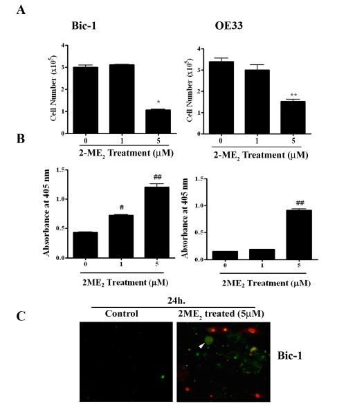
Figure 1. Antiproliferative and apoptotic effects of 2-Methoxyestradiol (2-ME2) on BEAC cells.
A. Bic-1 and OE33 cells were incubated with various concentrations of 2-ME2 for 24h or left untreated. Cell numbers were counted electronically using Cello meter Auto T4 Cell Counter. Data is displayed as mean ± SD in each case. P value was determined by Student’s t-test. *p<0.05 vs controls and **p<0.001 vs controls.
B. Bic-1 and OE33 cells were incubated with various concentrations of 2-ME2 for 24 h and apoptotic cell death was determined using cell-death detection ELISA kit. Data is displayed as mean ± SD in each case. P value was determined by Student’s t-test. #p<0.01 vs controls and ##p<0.001 vs controls.
C. Immunofluorescence images of Bic-1 cells treated with or without 5 μM of 2-ME2 for 24 h. Cells that have bound Annexin V-FITC show green staining in the plasma membrane, which signify early apoptosis. Cells that have lost membrane integrity will show red staining (PI) throughout the nucleus and a halo of green staining (FITC) on the cell surface (arrow head), while FITC Annexin V negative and PI positive (red) indicate late stage apoptosis and death. Viable cells are negative for FITC Annexin V binding and PI staining (control).

