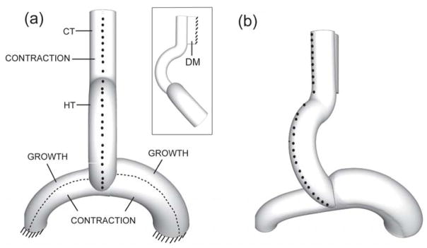
Figure 3.
Three-dimensional finite element model for looping heart without splanchnopleure (ventral view). Nodes on the ventral midline are marked to visualize rotation. (a) Undeformed configuration with morphogenetic loads indicated; insert shows side view. (HT = heart tube, CT = conotruncus, DM = dorsal mesocardium) (b) Deformed configuration. Midline nodes move rightward as HT rotates, similar to experiment (see Fig. 2B). (from 24)

