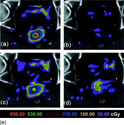
Figure 6.
The dose mapping inconsistency for the B-Spline DVF for a prostate image displayed on a single transverse slice. (a) and (b) show the dose consistency errors for DVF-only and PIDVF-with-DVF, respectively, for a 2×2×2 mm3 dose grid resolution. (d) and (e) show the DVF-only and PIDVF-with-DVF dose consistency errors when the dose grid resolution is 4×4×4 mm3.

