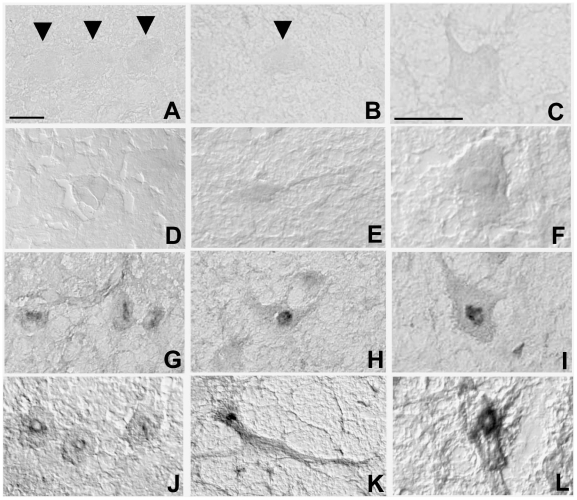
Figure 1. Reactivity of rat brain sections with serum IgAs and IgGs of CD patients.
Immunohistochemical staining on rat Purkinje cells (left panels, arrow heads), deep cerebellar nuclei neurons (middle panels, arrow head) and brainstem reticular neurons (right panels) using either serum from a healthy donor (A–F) or from an untreated CD patient (G–L). No binding of serum IgAs (A–C) and IgGs (D–F) from healthy donors could be detected. In contrast, antibodies in sera of CD patient (positive for anti-TG2 IgA/IgG antibodies) stained the cytoplasm and the perinuclear zone of neurons from the three brain areas, both when detecting IgAs (G–I) or IgGs (J–L). Calibration bars = 20 µm.

