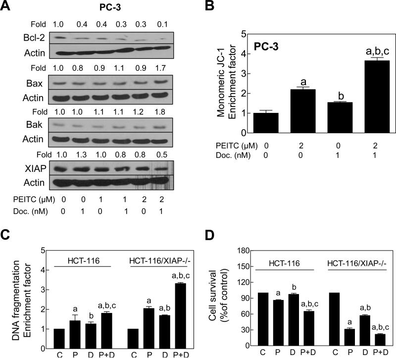
Figure 3.
XIAP protected against PEITC-mediated sensitization to Docetaxel. A,Immunoblotting for Bcl-2, Bax, Bak, and XIAP proteins using lysates from PC-3 cells treated for 24 h with PEITC and/or Docetaxel. Numbers on top of the bands represent change in protein level relative to DMSO-treated control (first lane). B, Flow cytometric analysis of mitochondrial membrane potential (monomeric JC-1 associated fluorescence) in PC-3 cells treated for 6 h with 2 μM PEITC and/or 1 nM Docetaxel (mean ± SE, n= 3). C, Cytoplasmic histone-associated DNA fragmentation, and D, cell viability in parental HCT-116 cells and its XIAP−/− variant (HCT-116/XIAP−/−) following 24 h treatment with DMSO, 2 μM PEITC (P), 1 nM Docetaxel (D), or the combination of PEITC and Docetaxel (P+D) Results shown are mean ± SE (n= 3). Significantly different (P<0.05) compared with aDMSO-treated control, bPEITC, and cDocetaxel by one-way ANOVA followed by Bonferroni's test.

