Figure 5.
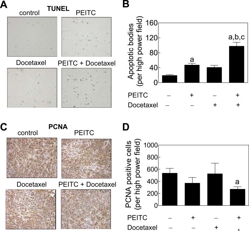
Apoptosis induction and proliferating cell nuclear antigen (PCNA) expression in the tumor sections. Microscopic images depicting A, TUNEL-positive apoptotic bodies, and C, PCNA expression in representative tumor section of the indicated group. Quantitation of B,TUNEL-positive apoptotic bodies/high power field, and D, PCNA expression/high power field in tumor sections from control, and PEITC and/or Docetaxel-treated mice. Results shown are mean ± SE (n= 3). Significantly different (P<0.001 for panel B and P<0.05 for panel D) compared with acontrol, bPEITC alone, and cDocetaxel alone group. Tumor sections from 3 individual mice of each group were examined.

