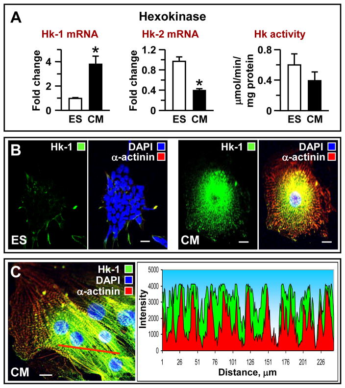
Figure 2. Transcriptomic and topological restructuring of the entry step in the glycolytic pathway: increase connection with mitochondria.
(A) A shift in hexokinase isoforms from Hxk-2 towards Hxk-1 and marginal reduction in enzyme activity in ES cell-derived cardiomyocytes (CM). Immunocytochemistry indicates that, compared to (B) ES cells, (C) cardiomyocytes have increased Hxk-1 abundance with a stippled pattern of intracellular localization and higher perinuclear concentration corresponding to mitochondrial network arrangement; the myofibrillar mesh is stained with α-actinin (red). Scale bar indicates 10 μm.

